How to prepare for polyp removal. Effective and safe ways to remove polyps in the uterus. How is the operation performed?
When polyps are found in the uterine cavity, they are removed. Previously, only the curettage method using vacuum aspiration was used. In advanced cases, open surgery was required. Nowadays it is most often used. This is a minimally invasive method of removing growths. Hysteroscopy is followed by a two-week recovery period.
Brief description of the operation
Hysteroscopy of the uterus is a minimally invasive technique. The operation is performed using a special hysteroscope. It is equipped with a mini-camera, with which an image of the mucous membrane of the organ is displayed on the screen. A hysteroscope has a hollow tube into which surgical instruments are inserted for manipulation. The method has a number of advantages over other methods of polyp removal:
- targeting of actions;
- minimal risk of relapse;
- has no age restrictions for mature women;
- does not interfere with conception and childbirth;
- minimal tissue damage;
- speed of operation;
- the procedure can be performed on an outpatient basis;
- minimal risk of complications (mostly their complete absence);
- no long sick leave is required.
Hysteroscopy varies by type. Depending on the equipment it is divided into:
- Radiosurgery. The formation is evaporated using radio waves.
- Mechanical. The polyp is excised or unscrewed with surgical instruments.
- Electrocoagulation. The polyp is separated from the mucous membrane using a loop-shaped electrode. Under current, soft tissue melts.
- Laser. The polyp evaporates quickly, leaving no scars.
Standard hysteroscopy of the uterus is done without hospitalization, without painkillers, under short-term anesthesia. If the polyp is large and the affected area is large or there are additional pathologies (endometriosis, fibroids, bleeding disorders, etc.) this is considered a complex case. The operation is performed in a hospital after the patient has been given general anesthesia.
Depending on the means administered orally to straighten the uterine walls, hysteroscopy is divided into gas (with carbon dioxide) and liquid (with physiological or 5 percent glucose solution).
Prescriptions and prohibitions for surgery
Hysteroscopy of the uterus is indicated for irregularities in the menstrual cycle, bleeding or discharge (including if they are caused by polyps). The operation is prescribed if:
- intrauterine synechiae;
- myomatous node;
- cancer affecting the mucous membrane or cervix;
- polyposis;
- not bearing;
- adenomyosis;
- abnormal development;
- infertility;
- perforation of the uterine cavity;
- foreign bodies;
- remnants of the membrane from the fertilized egg.
Hysteroscopy is also performed to examine the uterus before surgery, evaluate and monitor treatment. The procedure is mandatory when preparing for IVF or after a complicated birth.
Contraindications
Prohibitions include acute infectious pathologies, inflammation of the mucous membrane, and exacerbation of chronic diseases. The operation is not performed during pregnancy (including ectopic), cervical cancer, cervical atresia or bleeding. prohibited for kidney, liver, and cardiovascular pathologies.
Preparation and progress of the operation
The operation requires preliminary preparation. First, the patient donates blood and urine, and biochemistry is done. Smears are taken from the cervix and vagina. The following hardware diagnostic methods are used:
- fluorography;
The doctor is notified in advance about the presence of chronic diseases and allergies to specific drugs. If colpitis is detected, vaginal sanitation is done. Douching is stopped 7 days before surgery, and sexual intercourse is stopped 3 days before surgery. A woman should not use vaginal suppositories for at least a week before hysteroscopy of the uterus.
Before the operation, an enema is performed and the bladder is emptied. Immediately on the day of the procedure, patients are prohibited from eating. The operation is performed 5-7 days after the end of menstruation, when the mucous membrane has not yet recovered and the surface of the uterus is exposed.
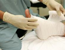
How is a polyp removed?
The patient is immersed in anesthesia, the genitals (externally and internally) are treated with an antiseptic. Then, with the help of gas or liquid, the uterine cavity is expanded and a hysteroscope is inserted into it. The organ, its mucous membrane, and the area occupied by polyps are assessed. If foreign fragments are present, they are pulled out using clamps that are inserted through the hysteroscope tube. Tissues are taken for analysis in the same way. Polyps are removed using special instruments. Then the cavity is disinfected and the hysteroscope is removed. The patient is gradually brought out of anesthesia.

Possible complications
Complications after the procedure include pain in the abdomen (inside) of a pulling nature. This is a natural reaction of the human body to surgery, as well as small discharges of blood without a strong odor. Due to the administration of anesthesia and the injection of air into the cavity, slight flatulence may occur. Sometimes short-term pain in the shoulders appears.
Serious consequences include persistent severe pain that cannot be relieved even with painkillers, or copious discharge of mucus and blood that has a putrid odor. The complete absence of discharge is also considered a pathological sign.
When removing polyps, soft tissues or the uterine cervix may be injured, and vascular hemolysis may occur. A very rare complication is an air embolism, when bubbles enter the blood. This may lead to the death of the patient. Sometimes chronic diseases worsen, defects form in the uterine cavity, hydrosalpinxes. However, any complications occur extremely rarely.
Postoperative recovery
During the rehabilitation period, to avoid the negative consequences of surgery:
| Prohibited | Recommended |
| · do not have sexual intercourse before your first period (at least 2 weeks) to avoid infection; · use of tampons; · douching; · lift weights and play sports for the first 30 days; · hot baths, baths, saunas, swimming in ponds; Ignore the urge to urinate; · use spermicides during the first 30 days. |
· after the procedure, use only sanitary pads; · You need to measure your temperature every day; · It is advisable not to take painkillers and aspirin (they thin the blood); · adhere to a proper diet to avoid stool upset. |
Immediately after hysteroscopy, the woman is given antibiotics. This will prevent possible infection, since damage to the endometrium can cause active growth of pathogenic bacteria. Antispasmodics help reduce negative symptoms and are prescribed immediately after surgical hysteroscopy.
To restore hormonal levels, appropriate medications are prescribed. They are aimed at stabilizing progesterone levels and reducing estrogen. If their balance is not maintained, then a relapse of the disease may occur. Also, hormonal therapy is aimed at preventing pregnancy, since conceiving a child in the first three months after hysteroscopy of the uterus is undesirable.
If after this no deviations from normal values are observed, then a spiral with gestagens can be implanted. This will allow you to quickly restore the damaged endometrium and avoid conception.
Oral contraceptives and gonadotropins are also indicated to prevent relapse. Therapy is carried out after checking hormonal levels or when distant formations are combined with other diseases (for example, adenomyosis, hyperplasia, etc.).
Herbal medicine is used as an auxiliary area. Decoctions, herbs or ready-made preparations can be used, but only after consulting a doctor. Self-medication is unacceptable, as it can cause some complications.
Recovery time
After surgical hysteroscopy, the damaged area is inflamed for the first three days. This is accompanied by an increase in temperature. Then healing begins. First, the wound closes with a crust, then falls off and the endometrium begins to grow. As a result, complete healing occurs in a couple of weeks. The recovery time depends on the size of the removed polyp.
As a result of electrocoagulation, the recovery period is up to 10 days. After other operations, wounds heal within five days and a little longer if the polyps were large. When using any type of hysteroscopy, the period of early rehabilitation is considered to be 4-6 weeks, and the final rehabilitation period is six months.
Histology
Immediately after hysteroscopy, the tumor and soft tissue obtained during curettage are sent for histology. Results are prepared within 10 days. The malignancy or benignity of the neoplasm and the risk of its degeneration into cancer are determined. Women over 50 years of age are at risk for developing an adenomatous tumor. This is a precancerous condition. Based on the results of histology, further treatment is prescribed if necessary. If cancer cells are detected, the patient is referred to an oncologist. Surgery may be done to remove the affected area.
Planning a pregnancy
Pregnancy after hysteroscopy can be planned only after six months. At this time, the menstrual cycle and the condition of the genital organs are monitored monthly. A woman should be regularly examined for sexually transmitted diseases, infections, and inflammations.
If any pathologies are detected, they must be treated before the child is conceived. If you plan to do IVF, then fertilization of the egg takes place at least six months after surgery. After the egg has been implanted, regular monitoring is necessary.
The rehabilitation period is always individual. Recovery depends on many factors. Mainly, it depends on the qualifications of the surgeons and their experience in performing such procedures. However, it is also important that after hysteroscopy, patients must follow all recommendations. This will not only speed up recovery, but also help avoid complications.
An endometrial polyp is a benign neoplasm of the uterine lining. The exact causes of endometrial polyp are still under study. In most cases, endometrial polyps occur as a result of hormonal disorders. However, there are cases when the appearance of a polyp is associated with trauma to the uterus; polyps appear after abortions, frequent curettages and other surgical interventions on the uterine cavity. The presence of an endometrial polyp is not considered dangerous to the life and health of a woman, however, it requires dynamic monitoring and treatment. There are several ways to remove an endometrial polyp: hysteroscopy, laser removal and diagnostic curettage.
What is an endometrial polyp
An endometrial polyp is a round-shaped formation that is attached to the uterus with a stalk. The stalk of the polyp contains many blood vessels that feed it. An endometrial polyp is formed as a result of local proliferation of the mucous layer of the uterus. The polyp is similar in structure to the endometrium and consists of glands and connective tissue.
Polyps can form as a result of hormonal disorders, which are characterized by excess production of estrogen and lack of progesterone. In the presence of hormonal disorders, pathological proliferation of endometrial cells occurs, which is accompanied by the formation of polyps.
Polyps can be single or multiple. They can be located in any part of the uterus: on its bottom, walls, corners and cervical canal. The size of polyps varies from a few millimeters to centimeters.
Endometrial polyps are often the cause of infertility. Polyps prevent the normal implantation of a fertilized egg into the endometrium. Lack of progesterone, as well as disturbances in the structure of the endometrium, lead to the risk of frequent miscarriages. Hormonal imbalance can be accompanied by anovulatory cycles, which also prevents conception.
In most cases, endometrial polyps do not threaten a woman's life. However, if the structure of the glands changes or the presence of atypical cells, the polyp can develop into a malignant formation. That is why it requires careful diagnosis and timely treatment.
Types of endometrial polyps
Endometrial polyps differ not only in location and size, but also in structure. There are four main types of endometrial polyps:
- Glandular polyp. It consists of overgrown endometrial gland cells. Glandular polyps are usually small in size (up to 1.5 cm). Most often, endometrial polyp of this type is not removed if its size does not exceed the permissible limit. Glandular polyps of the endometrium require monitoring over time, and can be removed on their own with the next menstruation.
- Glandular fibrous polyp. A glandular-fibrous polyp consists of endometrial gland cells and connective (fibrous) tissue. The size of this type of polyp can reach 2-2.5 cm. Their appearance indicates the development of a hormonal disorder. Removal of this type of endometrial polyp subsequently requires hormonal therapy.
- Fibrous polyp. Mainly represented by connective tissue. It has a denser structure and can reach 4-5 cm. A fibrous polyp must be removed.
- Adenomatous polyp. This type of endometrial polyp poses the greatest threat because its cells have an atypical structure. Adenomatous polyp must be removed.
Preparing to remove an endometrial polyp in the uterus
Removal of an endometrial polyp, regardless of the method, occurs under general anesthesia. Therefore, before the operation, the patient is prohibited from eating for 6-8 hours, and drinking for 2 hours. The patient must inform the attending physician about the presence of any allergies to medications.
Before the operation, the patient undergoes a coagulogram - a blood test for clotting. A woman may also be given an ECG (electrocardiogram) to check the condition of her heart.
Additional examinations include tests for infectious and viral diseases, in the presence of which the operation is prohibited (HIV, syphilis, chlamydia, etc.).
Removal of an endometrial polyp will not be performed if a woman is sick with influenza, ARVI and other infectious diseases. The procedure will be postponed until the woman has fully recovered.
After the endometrial polyp is removed, the woman will be able to go home on the same day, if there are no contraindications to this.
Endometrial polyp removal: curettage
Diagnostic curettage is the least effective method for removing endometrial polyps. Diagnostic curettage is almost always accompanied by a relapse of the disease, since the polyp is not completely removed, because the doctor acts blindly. In modern medical practice, this method is used less and less, since more effective methods for removing endometrial polyps have already been developed. Diagnostic curettage can be performed in emergency cases in the presence of significant bleeding, which is caused by hyperplasia or polyp in the endometrium.
Technique for diagnostic curettage:
- The cervix is dilated with special instruments.
- A metal loop, a curette, is inserted into the uterine cavity, which is used to remove the endometrium first from the bottom of the uterus, then from its walls and corners.
- The contents of the uterus are sent for histological examination.
There is also a separate diagnostic curettage, in which the mucous layer of the cervix is first removed. The contents are placed in one container. Next, the mucous layer of the uterus itself is removed. The removed endometrium is placed in another container. Thus, the uterine mucosa is analyzed from different areas.
Endometrial polyp removal: hysteroscopy
Hysteroscopy of an endometrial polyp is considered the most effective method of treating the disease. Hysteroscopy of an endometrial polyp allows removal of the tumor and pathological endometrium. With the help of endometrial polyp hysteroscopy, the surgeon will be able to see polyps that were not noticed during examinations.
Hysteroscopy of an endometrial polyp is carried out using a special instrument - a hysteroscope. The hysteroscope has the form of a tube, at the end of which there is a light source and a microvideo camera. The video camera transmits the image to the monitor screen in the operating room, so the surgeon can see everything that is happening in real time. The hysteroscope also contains surgical manipulators that will be used to remove the endometrial polyp.
Technique for hysteroscopy of endometrial polyp:
- The cervix is spread apart with special instruments.
- Liquid or gas is injected into the uterine cavity to expand it. This makes it easier to move surgical instruments in such a small space. The fluid or gas will leave the uterus on its own through the cervical canal.
- Manipulators are used to cut or unscrew pedunculated polyps.
- The attachment site is cauterized with electric current or a chemical solution.
- The entire pathological endometrium is removed.
- The removed material is sent for histological examination.
After hysteroscopy, relapses are rare. The polyp may appear in another place, which will indicate the ineffectiveness of treatment of the cause of the disease.
Removal of endometrial polyp in the uterus with laser
Laser removal of endometrial polyps in the uterus is considered the most minimally traumatic treatment method. This method specifically removes polyps in the uterus without leaving scars. This is important for women planning a pregnancy in the future. During the procedure, the doctor controls the layer-by-layer penetration of the laser, preventing injury.
Endometrial polyp removed: what next?
When the endometrial polyp is removed, the woman continues treatment for the cause of the disease. If the cause is a hormonal imbalance, the patient is prescribed a course of drug therapy. It may include combined oral contraceptives, gestagens (synthetic progestins), and gonadotropin-releasing hormone analogues. The average course of drug therapy is 3-6 months. The field is monitored by treatment: ultrasound and hormone levels in the blood.
After the endometrial polyp is removed, it is sent for histological examination. It will determine what cells the polyp consists of. This is necessary to exclude atypia. If malignant degeneration is suspected, the woman will undergo additional examinations. In the future, the issue of removing the uterus will be decided to prevent the development of cancer.
Start your path to happiness - right now!
An endometrial polyp is one of the types of endometrial hyperplasia, that is, the growth of its inner layer. The formation cells can gradually accumulate changes that are regarded as precancer, and then transform into endometrial cancer. So the uterine polyp itself is not a precancerous disease, but it is a precancer.
The most accurate method of diagnosing the disease is during which a biopsy of the polyp is performed, and subsequently its histological examination, that is, it is determined what cells and tissues it consists of. Any that was detected during hysteroscopy must be removed.
How to remove a uterine polyp
Many studies have proven that diagnostic curettage does not allow getting rid of these formations in all cases. Polyps consisting of dense tissues - muscle, fibrous (you can learn more about glandular-fibrous polyps from ours) are especially poorly removed in this way - the frequency of their disappearance after curettage is only 12%. Even simultaneous endoscopic control does not avoid relapses of the disease.
Effective removal of pathological tissue should affect the entire endometrium located under the formation, down to its deep basal layer. This can only be achieved by performing hysteroscopic intervention.
Methods for removing endometrial polyps involve the use of conventional hysteroscopic equipment, as well as the use of electrosurgical equipment or a laser guide. Removal of an endometrial polyp with a laser is a modern technology that allows you to completely get rid of unnecessary tissue, reduce the likelihood of bleeding from the removal point, and reduce the frequency of relapses. However, conventional hysteroresectoscopy, when properly prepared and performed, has very good results.
How to prepare for surgery

Before removing uterine polyps, the following diagnostic measures are carried out:
- examination of the cervix in mirrors, which helps to assess its condition, the shape of the cervical canal, the presence of an inflammatory process or damage to the organ; this is important because it is through the cervical canal that instruments for manipulation in the uterus will be inserted;
- bacteriological examination of smears from the surface of the cervix and vaginal walls to prove that the woman does not have bacterial inflammation of the genital organs, because otherwise there is a risk of infection in the uterus, which will cause endometritis;
- cytology smear;
- transvaginal ultrasound, in which a transducer is placed in the vagina and the uterus is examined without interference from the abdominal wall;
- general clinical examination - blood tests (general and biochemical) and urine, blood test for HIV, markers of viral hepatitis, electrocardiogram, chest fluorography, examination by a therapist.
Contraindications for polyp removal:
- inflammatory diseases of the vagina, cervix, uterus or appendages, caused by both common flora and sexually transmitted infections (for example) - surgery is performed after getting rid of these diseases;
- exacerbation of genital (thrush) or (vaginal dysbiosis);
- intense bleeding from the genital tract caused by endometrial hyperplasia or other causes until it stops;
- pregnancy;
- cervical pathology that prevents the passage of hysteroscopic instruments into the uterine cavity (cancer, stenosis, gross cicatricial deformation after ruptures during childbirth, and so on);
- severe concomitant diseases in the stage of decompensation (for example, diabetes mellitus with high levels of blood glucose and glycated hemoglobin, arterial hypertension with high blood pressure numbers) or exacerbation (for example, gastric ulcer, bronchial asthma and others);
- acute respiratory infection.
No special preparation is required for endometrial polyp removal. During the week before the procedure, sexual rest or the use of a condom is desirable. It is better not to use douches, vaginal tablets, suppositories and creams for any purpose.
On the day before the operation, you can take easily digestible food for lunch, excluding brown bread, cabbage, legumes, and it is better to refuse dinner or drink a glass of kefir. Liquid is not limited. On the morning of the operation, you should not eat breakfast or drink. In the evening and in the morning, a cleansing enema is performed as prescribed by the doctor.
The appropriate time for the operation is determined by the doctor, usually 2-3 days after the end of menstruation, that is, days 6-9 of the cycle, since at this time the endometrium has not yet recovered, but its menstrual rejection has already been completed. These days, polyps are better visible, they are easier to remove, and the operation is less likely to be accompanied by complications, such as bleeding.
Surgery

Surgery to remove an endometrial polyp is usually performed in a hospital. Hospitalization periods are short, do not exceed several days.
The patient is placed on a gynecological chair, and painkillers are administered intravenously. At the same time, the woman falls asleep and does not feel anything. General intravenous anesthesia can be replaced by spinal anesthesia or even endotracheal anesthesia. The decision on the type of anesthesia is made by the anesthesiologist depending on many factors, including:
- the likely duration of the manipulation and its volume;
- accompanying illnesses;
- drug intolerance, cases of allergies to the administration of painkillers;
- the possibility of complications during the operation.
In any case, adequate pain relief is necessary, since when the cervical canal is expanded for the introduction of a hysteroscope, pain and other negative reactions may occur.
How is the operation performed?
After putting the patient under anesthesia, the gynecologist treats the external genitalia with an antiseptic solution and inserts cervical canal dilators - special instruments that “stretch” the canal to the required size for free insertion of the hysteroscope. The uterine cavity is filled with liquid or gas to straighten its walls.

An effective method for removing endometrial polyps is hysteroresectoscopy.
Single polyps with a clearly visible stalk are removed using scissors or forceps inserted through the hysteroscope channel. These instruments, under visual control (the hysteroscope is equipped with a miniature video camera that allows you to see the operation area), are passed to the stalk of the polyp and cut through it. This procedure can be performed using a resectoscope loop. Laser polyp removal is carried out in the same way. After removal, the intervention site is carefully examined again to ensure that there is no formation.
If the polyp is located near the mouth of the fallopian tubes, technical difficulties of the operation arise, because in this place the uterine wall is very thin, only 3-4 mm, and the risk of damage increases. Therefore, mechanical separation of the polyp is used, and electrical resection is most often abandoned.
Resectoscopy using a loop electrode (electrosurgical polypectomy) is more often used to remove large formations located near the uterine wall (parietal) that have a dense fibrous structure. The loop is brought to the polyp and cut off to the base. If the removal is carried out mechanically, then it is first unscrewed, and then the polyp stalk is additionally removed with scissors or forceps inserted through the hysteroscope. In this case, the cervical canal is dilated with Hegar dilators.
How long does the removal take? The time of intervention depends on the complexity of the operation, the size of the polyp, its location, the experience of the gynecologist and many other factors. On average, the manipulation takes about 30 minutes. In case of multiple formations, technical difficulties when inserting a hysteroscope or removing the formation itself, the intervention takes longer. The duration of anesthesia is also increased if necessary.
Period after surgery
Normally, within 2-3 days after removal of the endometrial polyp, the patient experiences discharge . They are scanty, “smearing” and go away on their own as soon as the removal site “heals”. The patient may experience minor discomfort in the lower abdomen and in the area of the external genitalia; this is not dangerous and is associated with restoration of the cervix.
If your stomach hurts after the intervention, the doctor will prescribe painkillers. You can use products in the form of rectal suppositories; they are safer and no less effective than regular painkillers.
If pain intensifies and bleeding increases, as well as if it lasts more than 5-6 days, you should immediately consult a doctor. Such signs indicate complications of the procedure.
Negative consequences of hysteroscopy and polyp removal:
- perforation (perforation) of the uterine wall;
- bleeding from the site of removal of the formation.
During the first 2-3 days, a woman's temperature may rise. Most often this is a consequence of an exacerbation of a chronic inflammatory process in the fallopian tubes. In addition, after the removal of multiple polyps in the uterine wall, aseptic (microbial-free) inflammation occurs - a natural reaction of the body aimed at restoring the integrity of the mucous membrane.
When complications occur, repeated hysteroscopy is often performed, as well as curettage of the uterine cavity, antibiotics, detoxification agents, and hormones are prescribed.
- sexual rest for a week while the cervix continues to recover;
- refusal to use vaginal tampons;
- You should not douche or use vaginal dosage forms without a doctor’s prescription.
What not to do during the first week after surgery:
- visit a sauna, bathhouse;
- take a hot bath;
- go to the pool or solarium;
- play sports, do hard physical work.
The main questions that arise in the long-term period after polyp removal

When will your period start?
Despite the removal of the formation, the woman’s hormonal levels are not disturbed, therefore, menstruation after removal of the endometrial polyp occurs on time, only a slight deviation in the timing of the onset of menstruation is possible . Heavy periods are a variant of the normal course of the recovery period. However, if they develop into uterine bleeding, you should urgently consult a doctor.
When can you get pregnant?
Pregnancy after removal of an endometrial polyp can occur already in the current cycle if hormonal therapy is not started. However, this is not a completely favorable development of events, because the woman needs rehabilitation for a full recovery.
The optimal period for the inner layer of the uterus to completely recover is 3 months. It is for this period that combined oral contraceptives are prescribed. Their cancellation causes the so-called rebound effect, as a result of which the likelihood of pregnancy increases. If endometrial polyps were the cause, it is at this time that the most favorable moment for conception occurs.
What treatment is prescribed after removal of an endometrial polyp?
The question of the advisability of prescribing hormonal drugs remains controversial. Some doctors believe that when removing a small polyp, drug therapy may not be necessary. Others argue that hormonal therapy is advisable because it helps restore normal endometrial function. Hormones are prescribed for functional glandular polyps, adenomatous formations, as well as for the combination of polyps with endometrial hyperplasia.
Usually combined or progestogens (Duphaston) are prescribed. The choice of drug, its dosage and duration of administration is determined by the doctor. Usually it is 3 months. Often, a woman is offered to install it with the content of levonorgestrel - “”. These measures, in addition to restoring endometrial function, are also aimed at planning pregnancy.
Clinical observation of a patient who has undergone polyp removal is carried out for a year.
I hardly eat meat, so I thought that the low hemoglobin level might be related to this. The therapist assured: “If you don’t eat as much as you need, go to the gynecologist.” I haven’t been to a female doctor for several years - it’s an unpleasant thing. I used to go like clockwork - once every six months, then I changed my place of residence, drowned in a new - happier - life - and somehow stopped thinking about it: nothing bothers me much - and okay. This is mistake number one: you have to walk, absolutely.
How to find a polyp
A woman may not be aware that she has endometrial polyps. Therefore, an accurate diagnosis can be made of the pelvic organs; it is done on the fifth or sixth day of the cycle. Polyps can grow throughout the entire uterine cavity and “come out” of the vagina, and only under this condition can they be detected during a routine visual examination. Polyp is an unpleasant word, it feels like it’s some kind of alien. "Why? What is this? What is he... it... doing there? - I'm terrified that I'm 40 years old and I know absolutely nothing about this. “Well, what does it do... grows. It needs to be deleted. Yes, under general anesthesia. No, it’s not scary,” the doctor calmly answers.
What is an endometrial polyp
An endometrial polyp occurs in almost every second woman, and while previously women over forty who gave birth were more susceptible to this disease, now polyps are found in both very young and older women.
The uterus is lined with mucous membrane - the endometrium, and it - if anything is wrong in the body - tends to grow. A polyp is a benign formation of the uterine mucosa that occurs for various reasons: hormonal imbalances (low, high estrogen), diabetes, obesity, abortion, intrauterine device, inflammatory processes, various allergic reactions, and so on. Polyps can grow against the background of other diseases of the reproductive system - endocervicitis. The polyp consists of the same tissues as the endometrium and can be glandular-fibrous, glandular-cystic, or mixed. Often a polyp is depicted as a kind of “mushroom on a stalk”, but it does not always have this shape - the base may not be thin, but wide. The size of the polyp can range from very small to serious, and polyps measuring 1.5-2 cm already cause a lot of problems - pain, while small and single polyps may not bother you at all. Until they grow up.
Why is a polyp dangerous?
The larger the polyp, the more the woman’s body suffers from it: long, very heavy menstruation, bleeding and discharge in the form of blood clots after menstruation and in women during the period. Accordingly, large blood losses lead to a decrease in hemoglobin levels, which do not have time to recover. Low hemoglobin is bad for many reasons, but the common point is premature aging of the body. A lack of iron in the blood is indicated by increased fatigue, deterioration of the condition of the nails (when nails break and peel, you should not take calcium, but donate blood for hemoglobin). There may be pain in the lower abdomen, especially during menstruation (no, painful menstruation is not normal, it’s not enough to drink weed and assume that it’s hereditary and nothing can be done).
But what is equally important is that the polyp affects the reproductive function of a woman. Depending on its location, it can simply “block” the entrance to the uterus. If the polyp is large, it prevents the fertilized egg from attaching to the wall of the uterus.
Can a benign polyp develop into a malignant neoplasm?
The likelihood of this is very low, which is good news, but the removed material is always taken for histological analysis to ensure its safety. But the polyp, rarely degenerating itself, can serve as a background for the development of other inflammatory and oncological diseases of the endometrium.
Operation
A modern way to remove an endometrial polyp is hysteroresectoscopy, when an endoscopic tube is inserted into the uterine cavity under general anesthesia. With the help of a special loop passed under the leg of the polyp, the formation is cut off and the site of germination is cauterized in order to prevent recurrence.
The operation is considered almost bloodless, does not last long, a few hours after it the patient is allowed to go home, if there are no complications - pressure surges, bleeding, weakness, and so on. In my case, they started at one o’clock in the afternoon, I “woke up” at half past three, another hour and a half was needed for an IV with an antibiotic, pain relief (my lower abdomen hurt, I was given one injection, and nothing else bothered me - neither that day nor the next ). At five in the evening, on my feet and feeling normal, I went home.
An error occurred while loading.
The next day
Normally there should be no bleeding - only small brown discharge. In any case, a doctor's examination is necessary. I had a slight fever - no more than 37.3 degrees, this is normal. After the operation, I had to take antibiotics, an antifungal drug and... biokefir for 5 days to restore the microflora. The critical days passed with a bang: painless and short-lived (5 days versus the previous 7-8). The hemoglobin level increased, but, however, with the intake of iron supplements. Let's see what will happen next.
One month after removal
An ultrasound is done on the fifth or sixth day of the cycle, everything is fine, “the endometrium is thin, everything is clean. There are no traces." But the doctor was not only pleased with this: “If you want to go on maternity leave, work on it, it’s quite possible.” I don't want to yet, but knowing it's possible is great.
Prevention
The main thing is to visit a gynecologist and have a control ultrasound every six months. To prevent the appearance of endometrial polyps, it is necessary to control the body mass index, prevent inflammatory, autoimmune diseases and allergic reactions. For those who do not plan, it is possible to install a special intrauterine device with the drug. It both protects against unwanted conception and heals. The spiral is installed for five years and is not cheap - about 16,000 rubles. The feasibility is determined by the doctor: for example, it was not recommended to me, because the uterine cavity is deformed and there is a high probability that this expensive contraption will not stay there.
We thank Olga Terekhina, Ph.D., obstetrician-gynecologist at the Atlas Medical Center, for her assistance in preparing the publication.
Most experts are of the opinion that any tumor in the uterus must be removed. The same applies to polyps, which are round or mushroom-shaped formations on the mucous membrane of an organ. Of course, many patients are afraid to resort to radical treatment measures, especially if the polyps do not cause discomfort. To make an objective decision, let’s consider what risks exist in case of refusal of surgical intervention, and whether it is really necessary for small polyps.
The main reason for the appearance of polyps is hormonal imbalance, when a woman’s body experiences a lack of progesterone and an excess of estrogen.
The size of endometrial polyps in the uterus can reach 15 mm or more. If the diagnosed formation does not exceed 5-6 mm and does not cause discomfort, it is simply monitored. In some cases, polypous growths disappear on their own.
If it reaches 5-7 mm, it needs treatment. Some hormonal drugs reduce the growth of tumors and alleviate their symptoms.
In the absence of a clinical picture, close observation is possible. In case of increasing dynamics, surgical excision is performed.
Polyp sizes that exceed 13 mm are considered large.
They usually cause characteristic symptoms:
- pain in the groin;
- discomfort during sexual intercourse;
- bloody issues.
This clinical picture indicates the impressive size of the neoplasm, which is why it can be susceptible to regular mechanical irritation. In this case, surgery is mandatory.
Polypous growths may disappear on their own.
In any case, the decision to remove is made by the attending physician, who monitors the dynamics of progress.
In general, polypectomy is indicated in such cases:
- problems with conception or pregnancy;
- blockage of the fallopian tubes or cervical lumen;
- large polyps that cause discomfort, including heavy bleeding;
- the possibility of polyp degeneration into a malignant formation.
On what day of the cycle should I remove it?
Polyp removal is optimally carried out on days 6-9 of the menstrual cycle. During this period, the formations are better visible, since menstrual rejection has already ended, and the endometrium has not yet fully recovered.
How to prepare for surgery?
 Removing a polyp does not require any serious preparation, but there are some restrictions. A week before the procedure, it is recommended to refrain from intimacy.
Also undesirable:
- douching,
- candles,
- vaginal tablets,
- medicinal tampons.
On the day of surgery, breakfast and any kind of liquid are excluded.
What tests need to be taken?
Before surgery, each patient undergoes such diagnostic procedures:
- Examination on a gynecological chair using a mirror; the doctor evaluates the cervix for inflammation and other damage, examines the cervical canal through which instruments will be inserted during removal;
- Cytological examination of a smear to exclude malignant processes;
- Bacteriological examination of a smear to exclude a bacterial infection, because there is a risk of introducing it into the uterus, which may result in the development of endometritis;
- Transvaginal ultrasound, which reveals the location and size of polypous lesions;
- General clinical examination (general and biochemical blood test, urine test, electrocardiogram, fluorography, test for HIV and viral hepatitis, microreaction to syphilis).
Only after the results of all the above tests is the patient allowed to undergo surgery.
Removal and types of anesthesia
The procedure for removing polyp growths in the uterus is called polypectomy.
The operation is performed in several ways, depending on:
- type and size of formations,
- age and general condition of the patient.
The duration of removal depends on the complexity of the operation and can take from 20 minutes to several hours.
Polyp removal is optimally carried out on days 6-9 of the menstrual cycle.
In most cases, polypectomy is performed under general anesthesia; local anesthesia is indicated only for laser removal and in case of contraindications to general anesthesia.
Hysteroscopy
The advantage of this method is that the entire removal process occurs under supervision. To do this, they use a special device with a camera, which is inserted into the uterine cavity at the time of the operation and monitors the location of the polyp, as well as its size and shape.
Then, using a special tool, the polyp is cut out or “unscrewed”, and this entire process is displayed on the monitor.
The duration of hysteroscopy can take from 15 minutes to 2.5 hours, depending on the complexity of the surgical intervention.
Scraping
 This method of surgical intervention is considered less effective, since polyp removal occurs “by touch.” In this case, relapses are observed in 30% of cases.
The manipulation is performed under general anesthesia and only in case of contraindications is local anesthesia used.
An operation consists of the following processes:
- The patient is given a drug to expand the uterine walls.
- Anesthesia is administered.
- Dilation devices are inserted into the cervical canal.
- A carriage, a special instrument that resembles a spoon, is inserted into the organ cavity through the cervical canal, and the endometrium is scraped off with light pressing movements. Initially, the manipulation is carried out in the area of the polyp, then gradually moves to the internal os of the cervix, thus treating the entire cavity of the organ.
- The curettage site is treated with iodine.
If multiple polyps are found during surgery, they are initially removed, after which the entire uterine cavity is treated.
Scraping is done separately. First, they remove and then the growths in the uterine cavity.
After the operation, the patient undergoes a repeat hysteroscopy to exclude any remaining tumors. If they are detected, scraping is carried out again.
This method requires additional hormonal therapy to eliminate the risk of infection in the postoperative period.
Laser polypectomy
 Laser polypectomy is an innovative method for removing polyps. It allows you to avoid relapses and injury to endometrial tissue, unlike surgical intervention.
First of all, laser therapy is indicated for young and nulliparous women, for whom it is important to preserve reproductive function. Laser removal does not leave scars on the endometrial tissue, so the woman will not have problems conceiving in the future. However, this method is indicated only in mild cases when a “leg” is visible in the polyp.
The procedure is performed under local anesthesia. Depending on the size and type of polypous growth, a stronger or weaker laser is used.
Removal occurs by exposing the pathological tissue to a fixed laser beam, after which the area is promptly cauterized. The procedure takes 20-30 minutes.
Before removal, the patient undergoes the following measures::
- colposcopy;
- hysteroscopy.
Under the influence of a laser beam, a thorough, targeted removal of the pathological focus occurs without damaging healthy tissue. This allows you to avoid pain in the postoperative period and speed up the recovery process as much as possible.
Resectoscopy
This is another safe and effective way to remove polyps. Excision is carried out not only on the functional, but also on the basal layer of the uterus, which minimizes the occurrence of relapses.
After resectoscopy, recovery occurs quickly, but you will still need to take antibacterial drugs.
Like laser removal, resectoscopy specifically removes the formation without affecting healthy tissue. This method does not cause hormonal imbalance and does not affect the menstrual cycle, therefore it is used for women of reproductive age.
First of all, laser therapy is indicated for young and nulliparous women, for whom it is important to preserve reproductive function.
The procedure is performed under general anesthesia. A special device is inserted into the uterine cavity - an endoscope, at the end of which a camera is attached. Initially, the organ is diagnosed; if a polyp is detected, it is immediately excised using high-frequency current. In this case, all manipulations are displayed on the monitor screen.
After resectoscopy, women may experience nagging pain in the lower abdomen, which weakens on the second day and then completely disappears. In most cases, a hospital stay after the procedure is not required.
When is a hysterectomy required?
A hysterectomy is the complete removal of the uterus. It is carried out if cancer cells and multiple polypous growths are detected in the formation. It is extremely rarely used for women of reproductive age.
Unlike the methods described above for removing polyps, hysterectomy requires a long recovery under the supervision of medical personnel.
After the operation, the polyp tissue is sent for histology. Further treatment tactics will be determined based on the results of the study. Typically, hormone therapy is prescribed after a hysterectomy.
How long do you need to stay in the hospital after polyp removal?
 Depending on the method of polyp removal, with the exception of hysterectomy, the hospital stay after surgery takes no more than 3-4 hours.
Highly effective treatments such as laser polypectomy and hysteroscopy require only recovery from anesthesia.
During the next month after surgery, you must follow these recommendations::
- Avoid any physical activity;
- Take medications prescribed by your doctor;
- Avoid any vaginal interventions (tampons, suppositories, douching, intimate intimacy);
- Maintain intimate hygiene;
- Do not visit saunas, baths, swimming pools;
- Do not take a hot bath; it is recommended to take a shower with warm water;
- Do not lift objects heavier than 3-4 kilograms.
A month later, the inner surface of the uterus is restored and the endometrium is regenerated.
To speed up the process, the doctor may prescribe hormonal drugs that contain estrogens and gestagens, for example, Yarina. When removing a single polyp, additional therapy is usually not required.
Possible complications
In rare cases, complications such as these may occur after polyp removal:
- Recurrence of polyposis;
- Discomfort and pain during sexual intercourse;
- Long delay of menstruation (more than 3 months);
- Copious bleeding;
- Infectious lesion of the endometrium;
- Uterine perforation (can occur after blind curettage);
- Scarring and adhesions that make conception difficult or even lead to infertility;
- Development of cancer processes (these are extremely rare cases and occur due to incomplete removal of polypous growths).
After surgery, you must regularly visit your gynecologist. Any alarming symptoms should prompt an unscheduled visit to the doctor. If complications occur, the specialist will take the correct treatment tactics. Repeated hysteroscopy is often performed, then antibacterial, hormonal and detoxification agents are prescribed.
A month after the intervention, the inner surface of the uterus is restored and endometrial regeneration occurs.
It is important to know that in the first 2-3 days after surgery, a woman’s temperature may rise. There is no need to panic, since this is a natural exacerbation of the inflammatory process in the fallopian tubes.
Often, after removal of polyps, microbial-free inflammation is observed, which is also a natural reaction of the body aimed at restoring the mucous membrane.
An additional useful video about uterine curettage and possible complications after surgery:
Removal of polyps is a radical treatment method that many patients are afraid of. But innovative methods of polypectomy are nothing more than a quick and painless mini operation. However, for isolated small formations that do not cause discomfort, it is recommended to choose a wait-and-see approach. The most important thing for a woman is to find a specialist whom she can trust with her health.
You can make an appointment with a doctor directly on our website.
Be healthy and happy!